What Happens at the 3D Scan Appointment
The appointment where an implant case stops being a range and becomes a plan — about half a minute of imaging, an examination, and the decisions no flat X-ray can support, as Dallas–Fort Worth practices describe the visit.
Get an exact price for your implants.
A few quick questions, and one local practice sends you a written quote.
Five quick questions. Your answers go to one implant practice in your area — and your estimate comes back as an exact written quote from that practice, not a made-up range.
How this works
- Answer five questions — they tell the practice whether you need one implant or a full arch before anyone picks up a phone.
- One local practice gets your request. Not a marketplace, not a bidding list — one practice in your area.
- You get an exact written quote at a free consultation — implant, abutment, crown and fees in writing, before you decide anything.
Why no instant price?
Calculator sites invent a range from your clicks. We don't. The same mouth can price thousands apart depending on bone, extractions and the teeth you choose — so your estimate is a real quote from the practice that will do the work, with a 3D scan behind it. That is the number you can actually plan on.
Free · No obligation · We never sell your information. Your request goes to one local implant practice only.
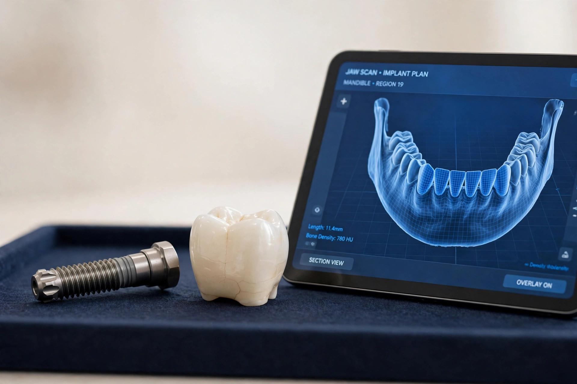
Every implant quote that is worth the paper it is printed on starts at the same appointment: the visit where a cone beam 3D scan turns your jaw from an estimate into a set of measurements. Dallas–Fort Worth practices describe that visit in their own patient pages, and the shape is consistent across the metro. This guide walks through what actually happens at the appointment, what the scan shows that a flat X-ray cannot, and which decisions are made from it before anyone schedules surgery.
What the scan itself is like
A cone beam scan (CBCT, for cone beam computed tomography) is a 3D X-ray of your teeth, jawbone, nerves and sinuses. You sit or stand with your chin resting on a small support, the imaging arm makes one slow rotation around your head, and the computer assembles a three-dimensional model you can be shown slice by slice. Published practice descriptions put the rotation at about 20 to 30 seconds. Nothing goes in your mouth, there is no film to bite on, and there is no injection or contrast dye.
A Dallas practice in Oak Lawn describes its version plainly: you stand still for about 30 seconds while its cone beam machine circles your head, and the practice gets a 3D view of your jawbone, nerves and sinuses. A Dallas oral surgery practice serving the metro describes the first visit the same way at the planning level — an exam, X-rays, and often a CBCT 3D scan to evaluate bone, nerves and sinuses, ending with a clear plan and cost estimate. The scan is the short part of the visit. The examination, the image review and the planning conversation are the appointment.
Why a flat X-ray is not enough
Planning an implant is a three-dimensional problem. The post has to live inside bone, with healthy bone on every side of it, the nerve that gives feeling to your lip and chin kept at a safe, measured distance below it, and the sinus floor respected above upper back teeth. A conventional X-ray shows a shadow of that anatomy: it can suggest height, but width — the dimension that decides whether a standard implant fits at all — is invisible on it.
The specialty radiology academy's position statement on implant imaging, published by the American Academy of Oral and Maxillofacial Radiology, recommends that cross-sectional imaging be used for the assessment of all dental implant sites, and that cone beam CT is the imaging method of choice for gaining that information. That recommendation is the reason the scan sits at the front of nearly every implant plan in this metro: the millimeters measured on the scan are the millimeters in your jaw.
What the scan shows the planning team
- Bone height, width and density at the exact site — whether there is enough bone to hold an implant securely, and how solid that bone is.
- The nerve canal in the lower jaw, followed along its whole path, so the plan can measure and keep its distance at the precise spot.
- The sinus floor in the upper jaw — how much room exists below it, and whether a sinus lift belongs in the plan.
- Problems a flat film hides — cysts, areas of infection, and old root fragments left from earlier work.
- The neighboring teeth and bite, so the implant is planned backward from where the finished tooth needs to stand.
The decisions that come out of the scan
A scan earns its place only if the plan is allowed to change because of it. The outcomes practices describe fall into a short list. The most common is to go ahead as planned, now knowing the numbers instead of estimating them. If width or height is short, the answer is graft first, then wait — the scan states precisely how much bone is needed and where. A different implant length or diameter can fit a site the first plan did not suit. Tilting an implant to work with the bone that exists, rather than building bone that does not, is a central idea behind All-on-4 style full-arch treatment.
The scan also feeds the surgery itself. Merging the 3D images with a digital impression of your teeth lets the planned position be built into a surgical guide — a template that sits over your teeth on surgery day and directs placement to the position, angle and depth chosen on the screen. Whether your case uses a guide is a planning choice your practice can explain from your own images.
Radiation, stated plainly
A cone beam scan involves more radiation than the small X-ray taken at a cleaning, and much less than a medical CT. A systematic review of cone beam imaging in implant dentistry reported effective doses from 5 to 652 microsieverts for limited-field scans and from 46 to 1,073 microsieverts for large-field scans. The wide range is the point: dose follows the size of the field scanned and the machine's settings, so the standing guidance is to scan the smallest field that answers the planning question, and to take the scan when there is a clinical reason — implant and surgical planning is the headline reason — rather than as a routine picture.
What the appointment costs in DFW, and what you leave with
Across this metro, the consultation pattern published by implant centers is a free consultation, with the 3D scan included at most dedicated implant centers and priced on its own — commonly in a $0–$300 band — where it is charged separately. Because practices bundle differently, the question to ask when you book is a simple one: is the scan inside the consultation price, or is it a separate line? Either way, it belongs as a named line in the written plan, not as a surprise.
What you leave with matters more than what the scan cost. A complete appointment ends with a proposed treatment sequence and a written estimate: what will be placed, what has to happen first if anything does, how long the stages run, and what each stage costs. If your consultation ends with a verbal number and no images reviewed with you, that is information about the practice. Take the written plan home, compare it against the published ranges in our Dallas–Fort Worth cost guide, and let the second practice you visit measure the same jaw.
The bottom line
The 3D scan appointment is where an implant case stops being a range and becomes a plan: about half a minute of imaging, an examination, and a set of decisions — proceed, graft first, resize, re-angle, guide the surgery — that no flat X-ray can support. Our form sends five answers to one local practice; the estimate comes back as an exact written quote after that examination and scan, which is the order the work actually happens in.
Common questions
How long does a 3D implant scan take?
The scan itself takes well under a minute. Published practice instructions describe the imaging arm making one slow rotation around your head — about 20 seconds at one practice, about 30 seconds at a Dallas practice with an in-office cone beam machine — while you rest your chin on a support and hold still. Nothing goes in your mouth, and there is no injection or dye. The longer part of the appointment is everything around the scan: the examination, the review of the images with you, and the written plan that comes out of them.
Why can't a regular X-ray plan my implant?
A flat X-ray is a shadow: it shows height, but not width. A ridge of bone can look tall and generous on a panoramic film and turn out, on a 3D scan, to be only a few millimeters wide. An implant has to sit inside bone with healthy bone on every side, at a measured distance from the nerve canal in the lower jaw and the sinus floor in the upper jaw. The American Academy of Oral and Maxillofacial Radiology recommends cross-sectional imaging for the assessment of dental implant sites and names cone beam CT as the imaging method of choice for getting that information.
How much radiation is in a cone beam scan?
More than a small dental X-ray, and far less than a medical CT — which is why the scan is used selectively, for surgical planning, rather than as a routine checkup image. A systematic review of cone beam imaging in implant dentistry reported effective doses ranging from 5 to 652 microsieverts for limited-field scans and 46 to 1,073 microsieverts for large-field scans; the dose grows with the size of the area scanned, so practices scan the region the plan actually needs. Your practice can tell you the field size and dose for your scan before it is taken.
What happens if the scan shows I do not have enough bone?
Then the scan has done its second job: finding that out before surgery instead of during it. The usual answers are a bone graft first, with the implant placed after the graft heals; a different implant size or angle that fits the bone you have — tilting implants to use existing bone is a central idea behind full-arch treatment; or, for some sites, a different treatment altogether. Each answer changes the timeline and the quote, which is exactly why the written plan comes after the scan, not before it.
Skip the guessing. Get the exact number.
Five questions, one local practice, one exact written quote — implant, abutment, crown and fees, in writing, at a free consultation.
Reveal My Estimate →